For patients
Understanding your patient-specific implant.
Your surgeon has told you about an implant designed for you. Here, in simple terms, is how it is designed and made, and what it changes in everyday life.
This information is general. Only your surgeon can answer your questions about your situation, your risks and your recovery.
The procedures
Rebuilding missing bone with a part made for you.
Cranioplasty
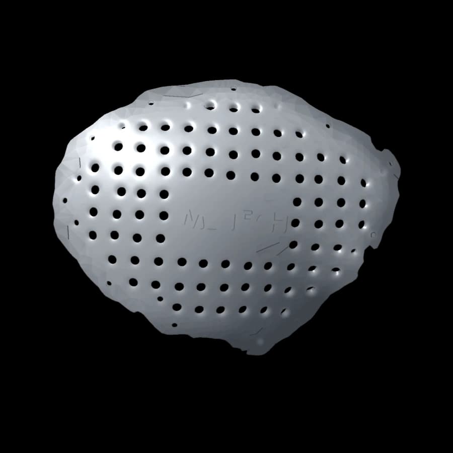
After an injury, a tumour or an operation that required removing part of the skull, cranioplasty closes this opening. It protects the brain and restores the shape of the head.
Orbital reconstruction
The orbit is the bony cavity around the eye. When its floor or rims are fractured or damaged, an implant restores the support of the eye and the contour of the cheekbone.
Why patient-specific
A shape decided before the operation.
A standard implant has to be cut and bent during surgery. A patient-specific implant is designed in advance on your CT scan: it matches the edges of the bone, and your surgeon approved its shape before it was made.
Fitted to your anatomy
Designed on your own bone, for precise contact with the edges of the defect.
A natural contour
The shape is rebuilt, often using the healthy side as a model.
Light and thin
A few grams for the orbit, about 140 to 160 g for a large cranial implant shown here.
Your journey
Six steps, from consultation to follow-up.
Timings depend on your situation and are set with your medical team.
01
Consultation
Your surgeon explains the operation, its benefits, its risks and the alternatives.
02
CT scan
An imaging scan of the head reproduces your anatomy in 3D. It is painless.
03
Design
The engineers design the implant on your anatomy. Your surgeon checks and approves the project.
04
Manufacturing
The implant is printed in titanium, inspected, then sent to the hospital where you will have surgery.
05
Surgery
The surgeon places the implant, designed in advance to fit your bone, and fixes it with small screws.
06
Follow-up
Your team schedules check-up appointments. Follow its advice on healing and returning to activity.
Titanium
A metal chosen for implants for decades.
Well tolerated
The titanium alloy used is a reference for implants in contact with bone.
Strong and light
Almost half the weight of steel, it allows thin, strong parts.
Corrosion-resistant
It remains stable in the body over the years.
Non-magnetic
Compatible with most imaging tests, including MRI under the specified conditions.
The implant is printed layer by layer: a laser melts a very fine titanium powder, following exactly the design approved by your surgeon. The part is then cleaned and inspected.
Technical data on the materialEveryday life
Living with an implant.
The answers below are general. Your surgeon’s instructions always come first.
MRI and CT scans
Titanium is not magnetic. An MRI is usually possible: always tell the imaging department about your implant, and they will check the scan conditions.
Security checks
Some sensitive scanners may detect the implant. Keep with you the document given to you after surgery.
Heat and cold
Titanium conducts heat poorly for a metal. Some people still notice extreme temperatures over the operated area.
Physical activity
You return to activity gradually, as advised by your surgeon. Ask them in particular before any contact sport.
Appearance
The implant sits under the skin and reproduces the contour of your skull or face. After a cranioplasty, hair grows back around the scar and usually hides it.
Your documents
Keep the documents given to you after surgery and tell your doctors and dentist about the implant.
Preparing your consultation
Questions to ask your surgeon.
Write them down before your appointment, along with your own questions.
- 1Why is a patient-specific implant suited to my situation?
- 2What are the other possible options, and their advantages?
- 3What are the risks of the operation and how are they monitored?
- 4How long is the hospital stay and the recovery?
- 5When can I go back to work, sport and driving?
- 6Which signs should make me seek medical advice quickly after the operation?
- 7Which check-ups and tests are planned afterwards?
Glossary
The words in your medical file.
- Cranioplasty
- An operation that rebuilds a missing part of the skull.
- Decompressive craniectomy
- Temporary removal of part of the skull bone to give the brain room when it swells.
- Orbital floor
- A thin bony wall under the eye that supports the eyeball.
- Zygoma (cheekbone)
- The cheekbone, which also forms the outer edge of the orbit.
- CT scan
- An X-ray imaging test that produces thin slices of the body.
- DICOM
- The standard format for medical images, used to send the CT scan.
- Segmentation
- The step that isolates bone on the CT images to build a 3D model.
- CAD
- Computer-aided design: drawing the implant in 3D.
- Additive manufacturing
- 3D printing: the part is built layer by layer.
- Laser fusion
- A metal printing process in which a laser melts a fine titanium powder.
- Ti-6Al-4V
- An alloy of titanium, aluminium and vanadium, widely used for implants.
- Patient-specific implant
- A device designed and made for one patient, from their anatomy.
A question about this technique?
Talk to your surgeon first: they decide on the treatment and send us your file. For a general question about our implants, you can also write to us.
Write to us